April 27, 2024, 07:25:11 AM
Forum Rules: Read This Before Posting


Topic: NMR CaM Binding  (Read 2600 times)

0 Members and 1 Guest are viewing this topic.

Offline burningdesire

  • Regular Member
  • ***
  • Posts: 10
  • Mole Snacks: +0/-0
NMR CaM Binding
« on: November 25, 2012, 10:41:35 AM »


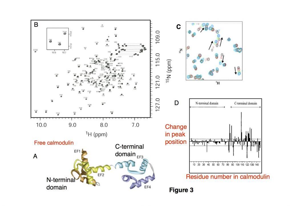
Calmodulin (CaM), a 16.7kDa EF-hand regulatory calcium-binding protein composed of two domains (Figure 3A) and mediates a range of cellular processes, undergoes a conformational change upon binding to Ca2+ ions, which enables it to bind to specific proteins. The resonances observed in the fingerprint of CaM have been assigned (Figure 3B) to their specific residue in the CaM sequence, which allows us to know which cross-peak in this 148 residue protein corresponds to an amino acid within the protein.

In this experiment, a 25 amino acid peptide substrate was bound to calmodulin in different ratios of CaM: peptide = 1:0, 1:0.5, 1:0.8, 1:1.2 and HSQC spectra were recorded for each sample (Fig 3C) (black= 1:0, red = 1:0.5, green 1:0.8 and cyan 1:1.2). 

We can measure the changes in the positions of the cross-peaks for each of the amino acids within the calmodulin during the titration and plot this (figure 3D)


Using these data and the figures, describe what structural changes occur in CaM when the peptide binds.  (5 marks total)

Offline Babcock_Hall

  • Chemist
  • Sr. Member
  • *
  • Posts: 5610
  • Mole Snacks: +321/-22
Re: NMR CaM Binding
« Reply #1 on: November 25, 2012, 04:18:13 PM »
Have you attempted an answer, and if so, what is it?

Offline burningdesire

  • Regular Member
  • ***
  • Posts: 10
  • Mole Snacks: +0/-0
Re: NMR CaM Binding
« Reply #2 on: November 25, 2012, 09:01:41 PM »
Have you attempted an answer, and if so, what is it?

I said C-terminus of CaM has great conformational change has the change in peak position is great.
I also said there are a lot of cyan dots in C, showing there with increased amount of peptide, the binding to CaM increases.

Not sure what else to say.

Offline Babcock_Hall

  • Chemist
  • Sr. Member
  • *
  • Posts: 5610
  • Mole Snacks: +321/-22
Re: NMR CaM Binding
« Reply #3 on: November 26, 2012, 09:11:20 AM »
With a little bit more information about how residue number relates to the structure, you might be able to say that a particular alpha helix or loop shows large chemical shift changes.  Also, it looks to me as if the system is in fast exchange on the NMR time scale, assuming that the various colors of peaks correspond to different concentrations of peptide.

Sponsored Links